پژوهشگران دانشگاه میلان، و دانشگاه برشیا ایتالیا در پژوهشی مشترک به بررسی نقش میزان بتاآمیلویید مغزی-نخاعی نشانگر زودهنگام تشخیص تصلبچندگانه پرداختند.
روش:
- در این پژوهش آزمایشی، ۴۰ بیمار که به تازگی تشخیص اسکلروز مالتیپل از نوع عودکننده-بهبودیابنده (RRMS) شرکت نمودند.
- سطح بتاآمیلویید مغزی-نخاعی در مایع مغزی-نخاعی تمامی شرکت کنندگان سنجش شد.
- بعنوان خط پایه، معاینه کامل عصبشناختی و تصویربرداری مغزی (MRI) از شرکت کنندگان به عمل آمد.
- ۲۹ نفر از شرکت کنندگان بعد از یکسال، بعنوان پیگیری مجدداً MRI مغزی و سنجش بتاآمیلویید را انجام دادند.
نتایج:
- در مقایسههای هر فرد، نشانگر زیستی بتاآمیلویید مغزی-نخاعی نوع بالا و نوع پایین هم در سنجش اولیه و هم در مقایسههای یکساله مخ باهم تفاوتهای معناداری دارند.
- همچنین، مقایسههای بین فردی بتاآمیلویید مغزی-نخاعی نوع بالا و نوع پایین نشان دادند که هم در سنجش اولیه و هم در مقایسههای یکساله مخ باهم تفاوتهای معناداری دارند.
- تحلیل رگرسیون چندگانه نشان داده میزان بتاآمیلویید مغزی-نخاعی هم بعنوان خط پایه و هم بعنوان پیگیری، نشانگری بسیار دقیق برای پیشبینی وضعیت بیماری اسکلروز مالتیپل هستند.
- هیچ ارتباط معناداری بین سطح تخریب کلی مغزی و سطح بتاآمیلویید مغزی نخاعی، یافت نشد.
- بنظر میرسد سطح تخریب مخچه در پیشبینی پیشآگهی ضعیف بیماری اسکلروز مالتیپل، بیش از تخریب مغزی، اهمیت دارد.
راهبردهای کارکردی:
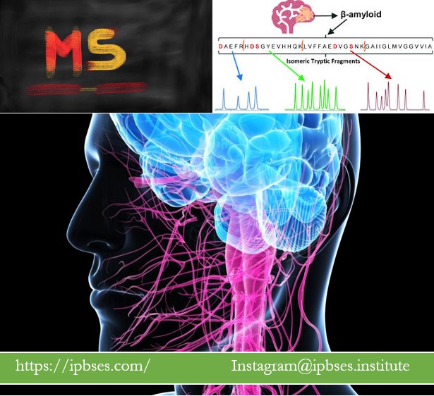
- آنالیز مایع مغزی-نخاعی برای سنجش سطح پروتیین بتاآمیلویید مغزی-نخاعی، روشی جدید، دقیق و سودمند برای پیشبینی زودهنگام وضعیت آتی بیماری در تصلبچندگانه است.
- در ارزیابی و سنجشهای MRI بیماران مبتلا به مالتپیل اسکلروزیس، بهتر است هنگام بررسی پاتوفیزیولژیک دستگاه اعصاب مرکزی (CNS)، توجه و تمرکز اصلی بر میزان تخریب عصبی مخچه باشد و میزان تخریب مغزی، بعنوان گام اول پیشآگهی در نظر گرفته نشود.
- سنجشهای مداوم MRI و بتاآمیلویید مغزی-نخاعی (و بطور کلی، مایع مغزی-نخاعی) بصورت حداقل سالانه، کمک شایانی به کنترل و مدیریت بیماری در مالتیپل اسکلروسیس خواهد نمود.
CSF β-amyloid predicts early cerebellar atrophy and is associated with a poor prognosis in multiple sclerosis
Abstract
Background
Neurodegeneration is present from the earliest stages of multiple sclerosis (MS) and is critically involved in MS related clinical disability.
Aim of the present study was to assess the connection between amyloid burden and early cerebellar grey matter (GM) atrophy compared to early brain GM atrophy in MS patients.
Methods
Forty newly diagnosed relapsing-remitting (RR-) MS patients were recruited. β-amyloid1-42 (Aβ) levels were determined in cerebrospinal fluid (CSF) samples from all subjects.
All participants underwent neurological examination and brain magnetic resonance imaging (MRI) at baseline. Twenty-nine out of 40 patients repeated a brain MRI at 1-year follow-up.
T1-weighted scans were segmented using the Voxel-Based Morphometry (VBM) protocol and the Spatially Unbiased Infratentorial Toolbox (SUIT) from Statistical Parametric Mapping (SPM12).
Results
Between-group comparison of cerebellar parenchymal fraction (GM+WM/total cerebellar volume%) showed significant differences between Aβhigh and Aβlow at baseline (p < ۰٫۰۰۰۱) and follow-up (p = ۰٫۰۲).
Similarly, a between-group comparison of cerebellar GM fraction (GMF) showed significant differences between Aβhigh and Aβlow at baseline (p = ۰٫۰۰۲) and follow-up (p = ۰٫۰۴).
The multiple regression analysis showed CSF Aβ concentration as the best predictor of GMF both at baseline and over time (β = ۰٫۵۰۵, β=۰٫۳۷۷; p < ۰٫۰۵).
No significant results were found regarding global brain atrophy and CSF Aβ concentration.
Conclusions
Early cerebellar atrophy seems to be crucial in predicting a poor prognosis in MS, more than early global brain atrophy.
Keywords
Multiple sclerosis, Cerebellum, β-amyloid, Progression
لینک منبع پیشنهادی برای مطالعه بیشتر  (further reading)

of the central nervous system (CNS) (Reich et al., 2018). Although its pathologic
hallmark is myelin loss, the neurodegenerative component is now considered remarkably
relevant, particularly as it is related to lon…
