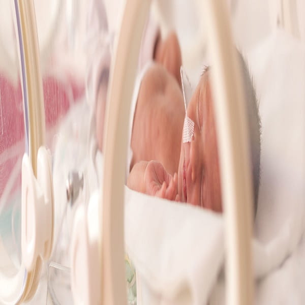
پژوهشگران دانشگاه ملی مستقل مکزیک در پژوهشی طولی در سطح ملی جامعه مکزیک به بررسی روش های نوین تشخیص و درمان آسیب مغزی مادرزادی با استفاده از متدهای پیشرفته نقشه مغزی، پرداختند.
روش پژوهش آسیب مغزی مادرزادی:
در این پژوهش طولی نوزادان دارای ریسک آسیب مغزی به مدت 8 سال در مکزیک مورد بررسی قرار گرفتند.
ابزارهای مورد استفاده شامل نوار مغزی QEEG، MRI، تست پاسخ فراخوانده شنوایی، تست پاسخ فراخوانده بینایی، ارزشیابی بیلی II بودند. از روش توانبخشی عصبی کاتونا هم به عنوان ابزار تشخیصی و هم ابزار درمانی استفاده شد.
یافته های پژوهش آسیب مغزی مادرزادی:
- بیش از 80% نوزادان دارای ریسک آسیب مغزی، ناهنجاری های قابل ملاحظه ای در MRI خود داشتند.
- 78% نوزادان زودرس دارای ریسک آسیب مغزی، در بررسی پیگیری 8 ساله رشد عصبی بهنجاری داشتند.
- تنها 69% نوزادان ترم دارای ریسک آسیب مغزی در پیگیری های 8 سال اول زندگی رشد عصبی بهنجار داشتند.
- آسیب های مغزی در نوزادان ترم به طور معمول بسیار شدید است.
- استفاده از روش های توانبخشی عصبی کمک شایانی به بهبود شاخص های رشد عصبی کودکان دارای آسیب مغزی مادرزادی می نماید.
راهبردهای کارکردی پژوهش آسیب مغزی مادرزادی:
- یکی از راهبردهای ایمن برای کاهش اختلالات عصبی، معاینات کامل عصبی از لحظه تولد نوزادان است.
- تمام کودکانی که از ابتدای تولد در بخش مراقبت های ویژه بستری می شوند لازم است مورد ارزیابی کامل عصبی قرار بگیرند.
- استفاده از شیوه های توانبخشی عصبی (همچون روش بیلی II) به کاهش دامنه آسیب های مغزی و بهبود رشد عصبی نوزادان دارای آسیب مغزی کمک زیادی می کند.
Early diagnosis and treatment of infants with prenatal and perinatal risk factors for brain damage at the neurodevelopmental research unit in Mexico
Abstract
Prenatal and perinatal risk factors for perinatal brain damage frequently produce brain injuries in preterm and term infants.
The early diagnosis and treatment of these infants, in the period of higher brain plasticity, may prevent the neurological and cognitive sequels that accompany these lesions.
The Neurodevelopmental Research Unit at the Institute of Neurobiology of the National Autonomous University of Mexico has taken this endeavor.
Method:
A multidisciplinary approach is followed. Pediatric, neurologic and rehabilitation clinical studies, MRI, EEG, visual and auditory evoked responses, and Bayley II evaluations are carried out initially.
Infants are followed up to 8 years, with periodic appointments for evaluation and treatment. Katona’s neurohabilitation method is used for initial diagnosis and treatment. Selective visual and auditory attention are explored from 3 months of age.
This method was created in the Unit and, if deficiencies are observed, the method also describes the treatment to avoid subsequent alterations of these processes.
Deficiencies in the acquisition of language are evaluated from 4 months of age, implementing treatment through instructions to parents on how they should teach their children to speak.
This method has also been developed in the Unit and is in its validation process. In the MRI, we pay special attention to subtle and diffuse patterns, due to the high frequency with which they appear in contemporary cohorts at a national and international level.
Results:
More than 80% of these infants showed abnormal MRI findings that should be taken into consideration.
The outcome of children at 8 years old showed that 78%, 76% and 78% of extremely preterm, very preterm and late preterm, respectively, had a normal neurodevelopment.
In term infants, only 69% had a normal neurodevelopment; in this group, the majority of infants had very severe brain lesions.
Conclusions:
It is necessary to evaluate, at an early age, all newborns with prenatal and perinatal risk factors for brain damage.
Special attention should be payed to all premature newborns and those newborns who have been discharged from the intensive care unit.
Keywords
Perinatal brain damage, Katona’s neurohabilitation, MRI Preterm Attention, Perinatal risk factors, Prenatal risk factors, Dr. Amir Mohammad Shahsavarani IPBSES Perinatal Brain mapping.


